Microscope endodontics — single-visit root canal with bioceramic obturation
Endodontics — root canal treatment — has the worst reputation of any dental procedure for reasons that are mostly historical. Done with a 1970s technique under loupes (or worse, naked eye), it was uncomfortable and unpredictable.
At HEC we perform endodontics as a 90-minute daycase appointment. No inpatient stay. The procedure is done in one sitting — we do not split a case across multiple visits without a clinical reason.
What it is
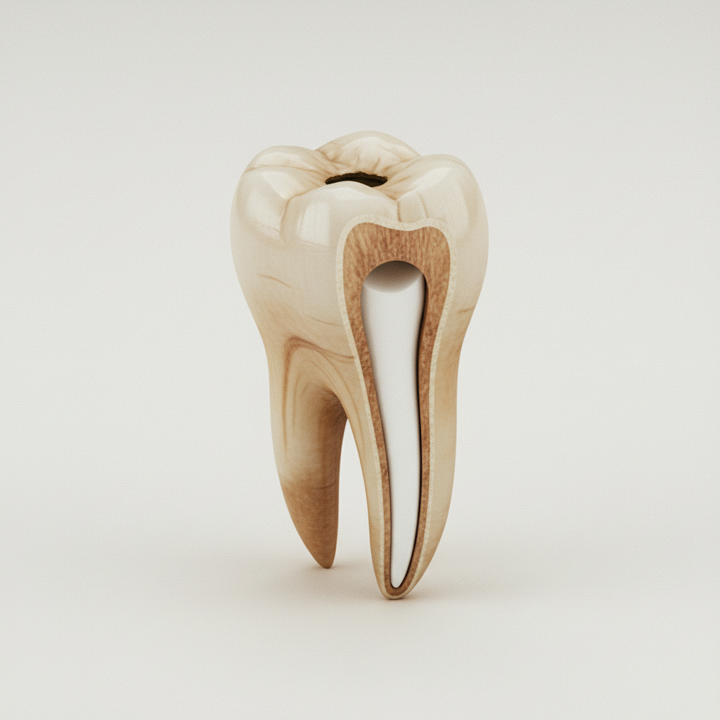
Root canal treatment removes the inflamed or infected dental pulp (the nerve and blood supply inside the tooth), shapes the resulting canal system, disinfects it, and seals it with an inert filling material. The tooth then receives a permanent restoration (typically a crown for posterior teeth, a build-up + composite for anterior teeth) to restore function and prevent fracture.
The procedure has three phases that the operating microscope makes visible:
- Access cavity preparation — a precisely positioned opening through the crown to reach the canal orifices. Under the microscope this is conservative; the goal is to find and instrument every canal while preserving as much sound tooth as possible.
- Canal shaping and disinfection — nickel-titanium rotary files mechanically clean and shape the canals to a calibrated taper; sodium hypochlorite + EDTA irrigation dissolves residual organic and inorganic debris.
- Obturation — the canal is sealed with a bioceramic sealer (Endosequence BC, AH Plus Bioceramic, MTA-based equivalents). Bioceramic sealers bond chemically to dentin and have superior long-term sealing compared with the older resin-based and zinc-oxide-eugenol materials.
Why microscope, why bioceramic — and why it matters
A standard endodontic procedure under loupes finds 70–80% of canals on first attempt. Under the operating microscope, with proper magnification and illumination, the find rate rises to 95%+. Missed canals are the single largest cause of endodontic failure.
Bioceramic obturation materials have a 5–10% better long-term seal than the older gutta-percha + AH Plus combination. The chemistry is more biocompatible with periapical tissues; the setting expansion (small but consistent) eliminates the micro-gaps that develop with eugenol-based sealers over time.
Together — microscope + bioceramic — these are the two upgrades that move 5-year success rates from the 85% range to the 92%+ range. We use both as standard.
Same-day choreography
| Time | What happens |
|---|---|
| 10:00 | Pre-op periapical radiograph, periodontal check, occlusal analysis |
| 10:15 | Local anaesthesia, rubber dam isolation |
| 10:30 | Access cavity preparation under the operating microscope |
| 11:00 | Canal shaping with NiTi rotary system; irrigation protocol |
| 11:45 | Final irrigation, drying, bioceramic obturation |
| 12:15 | Post-op periapical radiograph; coronal seal placed |
| 12:30 | Discharge with NSAID protocol and crown referral |
Anterior teeth (single-canal) typically finish in 60 minutes; multi-canal molars take the full 90 minutes. Re-treatments (failed previous endodontics) can extend to 120 minutes and may require a second short visit.
Who is a candidate
- Tooth restorable after endodontic treatment (sufficient sound tooth structure to support a crown)
- No vertical root fracture (immediate extraction indicated)
- No advanced periodontal involvement at the same site
- No untreated systemic infection that could complicate healing
Cases unsuitable for endodontics: vertical root fractures, teeth with insufficient remaining structure for crown placement, teeth with severe periodontal-endodontic combined lesions where extraction + implant is the better long-term answer. We say so in writing and recommend extraction or referral when this is the case.
What it treats
- Irreversible pulpitis (severe pulpal inflammation, often acute pain)
- Pulp necrosis with or without periapical lesion
- Periapical abscess
- Failed previous endodontics (re-treatment)
- Resorption defects (selected cases)
Recovery
Hour 0–6 — Local anaesthesia wears off; mild tenderness when biting on the treated tooth. NSAID (ibuprofen 600 mg every 8 hours) covers most patients.
Day 1–3 — Tenderness reduces; chew on the opposite side.
Day 7–14 — Most patients forget which tooth was treated.
Day 30 — Crown must be placed by this point. A treated tooth without a crown is at significant risk of fracture. We refer to a home dentist if you cannot return for crown placement at HEC.
What it is not
Endodontics is not a "second-best" alternative to extraction + implant. For a restorable tooth with healthy periodontal tissue, root canal + crown has comparable 10-year survival to single-tooth implants and preserves the natural tooth root, periodontal ligament, and proprioception that an implant cannot replicate. The decision is not "implant is better" — it is "what does this case need."
It is not painful in the modern protocol. The pain associated with endodontics in popular memory is the pain of pulpitis (the condition that brings patients to the chair), not the procedure itself. Under local anaesthesia with rubber dam isolation, the procedure is comfortable; post-op tenderness is mild and short.
It is not a substitute for the coronal restoration. A root-canal-treated tooth must receive a crown (posterior) or appropriate composite reinforcement (anterior) within 30 days. We say so explicitly; missing this step is the most common cause of treated-tooth fracture.
The package
The locked daycase package: pre-op radiograph, the procedure under operating microscope, bioceramic obturation, post-op radiograph, coronal seal, NSAID supply, optional one hotel night with transfers, translator, and the crown referral plan (HEC placement at follow-up or home-dentist referral with a written letter).